
Most people are probably familiar with ultrasound to some extent, physical therapists will often apply therapeutic ultrasound to injured tissues and ultrasound is typically used to visualize unborn babies. Musculoskeletal ultrasound is closer to the ultrasound utilized during pregnancy, the idea being that the probe emits super high frequency ultrasonic waves which go through a gel medium before being transmitted through the skin and different body tissues (such as muscles and tendons) before bouncing back and being received by the probe.
The frequency of these ultrasonic waves produces an image that is incredibly high resolution (3 times higher than MRI). The ability to view these images in real time is unique compared to other imaging modalities, allowing the doctors to observe the dynamic movement of body structures. An additional benefit of this is the ability for the patient to give feedback in real time, this is important because it is clinically significant whether or not the patient is experiencing any pain at the location of the probe where inconsistencies are observed. This can help the doctors discern whether the irregularities are concordant with their symptoms or simply incidental.
Patients will often come into our office and state that they have had a previous cortisone injection performed by another physician. While they probably did have a corticosteroid injected, that isn't really the relevant detail. More important is where the medication was placed, and how it got there. Historically, and the way that most physicians today do these injections is on a landmark basis. What that means is the doctor will insert the needle and once a landmark is reached (often when the needle hits a bone), then the placement of the needle will be adjusted relative to that landmark before the medication is injected. While injections done with this method can give patients relief, this technique doesn't completely take into account the variety in each person's anatomy or allow for the precision required to reliably hit the intended target. Even if the target is missed, it is likely that the medication was somewhat absorbed by being injected in the general area, giving the patient variable levels of relief.
Having the ability to use musculoskeletal ultrasound to guide these injections has been shown to dramatically increase the efficacy of these injections (check out these two articles). The ultrasound machine allows the target structure and the needle to be visualized in real time, which has multiple benefits. First, by seeing the needle in real time, landmarks do not need to be used, making the procedure more comfortable for the patient by allowing the physician to avoid having the needle touch down on bone, which can often be quite painful. Another advantage of using ultrasound is that the needle can be more precisely directed towards the target, allowing the doctor to know exactly where the medication is being injected. This can be helpful in a situation where the injection doesn't offer the patient any relief, allowing the doctor to definitively rule out that particular structure as the pain generator.
Let's use shoulder pain as a common example. The supraspinatus is a structure that can contribute to shoulder pain in many patients. One example of an injection where the benefit of ultrasound is obvious is a subacromial bursa injection where medication is injected into the subacromial bursa directly above the supraspinatus, bathing the tendon in medication. The placement of the medication is especially important because if the medication were to be injected directly into the tendon and there was even a small tear present, it could lead to even further degradation. The subacromial bursa itself is maybe a millimeter or two thick, smaller than the width of the average needle used to inject it, as you can see in the images below. You can see how even the best sports medicine physicians in the world would have trouble reliably hitting this target when using a landmark based technique.
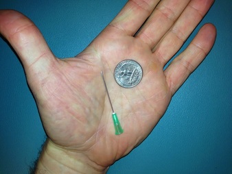
This is an width of the average needle used to inject medication. You can see how small it is.
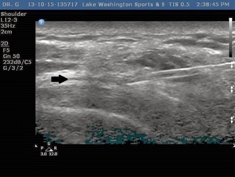
As you can see in this ultrasound image, the bursa (indicated by the arrow) is a very thin fluid filled sac smaller than the tip of the needle
No comments:
Post a Comment