
Joint inflammation may stem from inflammation of bursa(e), fluid filled sacs that provide cushion between bones, tendons, and/or muscles around a joint. They reduce friction and allow for free movement of surfaces moving in different directions. When inflamed, a normally slippery bursa becomes thickened and swollen, causing it to lose its ability to glide, which can become more irritated with movement. If you follow the Mariners, you may recall that this is a similar issue that afflicted top pitching prospect Taijuan Walker early on in spring training this year, preventing him from being able to compete for a spot in the starting rotation. While inflammation can be caused by an acute trauma such as a contusion, it is typically the result of a repetitive overuse injury caused by prolonged and excessive pressure. With a professional baseball pitcher, you can see how this could become an issue as their job description requires them to repetitively use their shoulder for prolonged periods of time. However, this is a very common injury that brings patients into our office all the time and can affect people of all ages and skill levels.
The general treatment plan for shoulder inflammation calls for ice and rest. While this can reliably help reduce the inflammation and thus pain level and discomfort, it doesn't change the underlying cause of the injury. Poor biomechanics often contribute. During Taijuan Walker's rehab, he worked on the mechanics of his pitch delivery. The can be deduced that the Mariners coaching staff and sports medicine team believed that there was a part of his delivery that was placing undue pressure on his shoulder which caused this inflammation in the first place. In fact it had been reported that he was complaining of shoulder soreness even before spring training started. But since finally coming back, armed with a refined delivery, he hasn't had any issues with his shoulder, recently striking out 13 batters in a start for AAA Tacoma.
The situation with Justin Verlander is slightly different. While the injuries are similar, from reports that I have seen the inflammation in Taijuan Walker's shoulder appears to be isolated to one particular area whereas the source of inflammation in Justin Verlander's shoulder was more complex. In addition, Taijuan Walker just recently turned 22, while Justin Verlander is 31 and pitching in his 10th major league season. Until this most recent incident, he hadn't missed a single game due to injury in his entire career. It would seem that if there were a biomechanical issue involving his delivery, this problem would have surfaced at some point in the past 10 years. A topic that will be covered in another blog post but that is relevant to this situation is injury recovery as we age. Being a pitcher in professional baseball takes an enormous physical toll; being asked to step on the mound every 5 days and torque various joints and limbs repeatedly (~100 times) in order to hurl a baseball at extreme velocities is no small task. Of all the pitchers in baseball, Justin Verlander has had one of the largest workloads, pitching over 200 innings in each of the past 7 years. As Justin Verlander ages, as with the recreational athlete, he is not going to be able to recover as quickly or efficiently as he once could. This case of shoulder inflammation could just be a case of father time finally catching up to him.
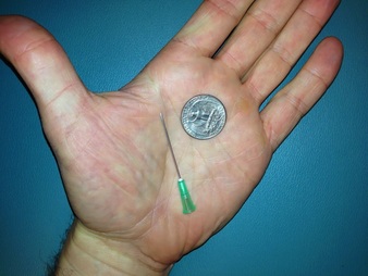
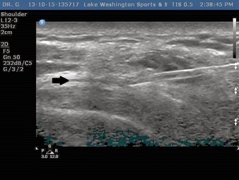
While Taijuan Walker seemed so have recovered fine, and Justin Verlander's recovery going well enough that he is tentatively scheduled to return to action this weekend, there are still instances where despite sufficient rest and biomechanical changes, the inflammation doesn't completely resolve and residual pain and discomfort remain. This is where a target intervention such as a corticosteroid injection can come into play and be quite helpful. It is widely believed that cortisone works in large part by by reducing inflammation. Injecting cortisone into the inflamed area, reduces inflammation and pain. One of the potential downsides is that corticosteroids, if accidentally injected into the body of a tendon can weaken that tendon, increasing the likelihood of rupture or tearing. Most of this risk is associated with the most common way of doing these injections, using palpation or landmark guidance. As discuss in our previous post on musculoskeletal ultrasound, when doing a landmark based injection, there is no way to know for sure where the medication actually ended up. As you can see in the picture below taken from that blog post, the size of the bursa (indicated by the black arrow) is tiny, especially compared to the needle being used to inject the medication. As you can imagine, the chances of being able to get the medication into that small space based on palpation alone is quite slim. Thus the risk of weakening the tendon by exposing them to corticosteroids is likely the result of missing the bursa and injecting the medication directly into the tendon. When patients come into our office we can visualize the inflamed bursa and get feedback from the patient about where their symptoms are coming from in real time. Then if the doctor determines that the patient would be a good candidate for an injection, the needle can be directed towards the target with certainty, and the medication can be seen while administered in real time.
