Thursday, April 23, 2015

Tennis elbow: Not just for tennis players!


Tennis elbow is a significant problem that can prevent people from performing athletically and occupationally.  Tennis elbow, or lateral epicondylopathy, is an overuse injury that presents as pain and tenderness on the outside of the elbow.  While 50 % of tennis players will develop tennis elbow during their lifetime, they represent only 5% of patients diagnosed with tennis elbow.  Plumbers, cooks, auto mechanics and other workers with occupations requiring repetitive gripping and grasping activities represent a large portion of people with LE.  People of all ages with overuse can acquire tennis elbow, but most cases of the injury occur in people between the ages of 30 and 50.


So, how do we treat tennis elbow?  Lateral epicondylopathy should first be treated with rest, ice and activity modification.  For more serious cases, prolotherapy and PRP are great options to amplify the body’s healing response.  It’s important to note that cortisone or “steroid” injections should not be used as a temporary quick-fix.  Patients often report feeling better at the time of the injection, but often end up with poor future outcomes likely related to a combination of tissue degradation from the cortisone and lack of biomechanical alterations (causing them to re-injure themselves).

A great tool to help see the lateral epicondyle is the use of musculoskeletal ultrasound.  Ultrasound is the imaging test of choice for the lateral epicondyle, for a reasons:
1. The highest level of resolution and detail

2. The level of detail is particularly excellent for tendon, ligament, and fascia, which are types of tissue that are not always seen clearly on other imaging tests (e.g., x-rays and MRIs)
3. It is the best test for checking motion
4. It is the only imaging test where we can also test for tenderness (since the ultrasound probe is touching the affected area)


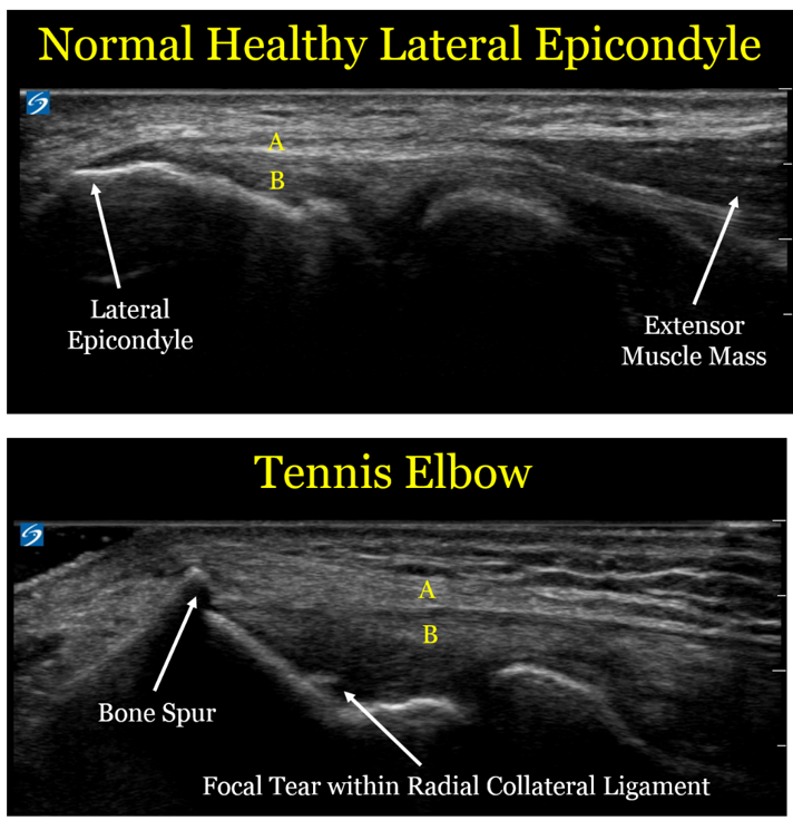
Here are comparison ultrasound images of a healthy lateral epicondyle, followed by the lateral epicondyle of a patient with tennis elbow. 


Let's look at the ultrasound images above, first focusing on the upper image of a healthy tendon.  One useful thing to see visually is that the muscle mass (the right arrow) looks distinctly different than the extensor tendon (designated by the yellow letter "A").  This is an important detail, since muscle has an excellent blood supply and heals well.  Tendon is made of a tight bundle of collagen with a relatively poor blood supply, and may struggle to heal.

In the second image, we can see the anatomic changes associated with tennis elbow:
1. The extensor tendon (A) is thickened and not as well defined as in the healthy elbow.  Of note, the patient with the healthy tendon is actually a much larger and more muscular person, so the thickened tendon is in fact a sign of an unhealthy tendon
2. The bone spur created by the unhealthy tendon.  We call this an enthesophyte, which is where hte tendon inserts onto the bone.  The reason these enthesophytes form is because bone is really a very dense putty, so the enthesophyte is caused by abnormal pulling forces by the unhealthy tendon
3. The part labeled "B" is the radial collateral ligament (RCL), which is underneath the extensor tendon.  In the case of the unhealthy patient, there is a tear in the RCL.  By definition, a sprain is a tear in a ligament.
4. Of note, there is no "inflammation", which is why the term -itis (e.g., tendonitis) is usually a misnomer.  This is an example of a chronic degradation of the extensor tendons and underlying radial collateral ligament, and the more proper term would be tendinosis or tendinopathy.


For a sake of comparison, the image below is an MRI


The level of detail is far less.  The resolution (which is defined as the ability to distinguish 2 points as distinct) is 5 times higher for the ultrasound.  In the ultrasound image, there is a clear distinction between the extensor tendons and the underlying radial collateral ligament, and we can see the individual fibers with great detail.  In the MRI, the white arrows show what is essentially a black line, with far less detail.  MRI does have some advantages in other contexts (e.g., it sees a wider field of view and can penetrate the bone, so it helps with bone contusions and certain types of joint injuries), but for imaging the lateral epicondyle, ultrasound is a better choice.


So what should we do with these patients?
- Historically, physicians have often performed landmark-based injections to the lateral epicondyle of cortisone.  However, moreso than for any other sports injury, repeated data show this is not a good idea.  While cortisone injections may help in the short term, they clearly make the injuries worse in the long term.
- Similarly, patients are often advised to use the non-steroidal anti-inflammatory drugs (NSAIDs) like ibuprofen or naproxen.  However, these are also bad choices, because while they can help with pain in the short term, they also prevent healthy collagen formation.
- The best initial treatment option is repeated icing.  Ice is a better anti-inflammatory than any medication, and also has other healing benefits.
- Physical therapy is also a mainstay of the best treatment for tennis elbow, where they can focus on exercise protocols designed to promote healing and fix damaging biomechanical patterns.
- For the subset of patients who do not improve with either ice or physical therapy, we often recommend use of proliferative therapy techniques including platelet-rich plasma injections or prolotherapy, which can help induce healthy healing.



Meet the Team: James Kim!


What is your role on the Team at Lake Washington Sports & Spine? 
 I am Dr. Chimes's new medical assistant. I will help patients on their path to recovery and will do whatever I can to Keep People Active! 


As the newest member of our team, we’d love for our patients to get to know you better.  Can you tell us 5 facts about James?
1) I was a Peace Corps volunteer in the country of Armenia from 2011-2013. Ask me anything about the country itself, what the Peace Corps was like, or how to live without indoor plumbing!
2) I'm a new transplant to Seattle! I have previously lived in New York, Illinois, and New Orleans. I miss Zabar's, Portillo's, and weekly parades/festivals. 
3) I used to work in bicycle shops as a mechanic. Got bike issues? Let me know. 
4) The first time I ate raw cookie dough was in 2012.
5) I cannot, for the life of me, figure out how to hula hoop. 


When did you first become interested in medicine (or why is the field of sports medicine appealing to you)?
As a child, I was encouraged (read: pushed) into studying for a career in either law or medicine. I was naturally inclined towards math and science, and liked figuring out how things work and how to fix them, so medicine seemed the right choice. Besides, I was never very good at arguing. Sports medicine appeals to me because I've always watched sports, as I grew up in Illinois during peak sports years, i.e. the Michael Jordan years. I'm also quite active myself, and am continually fascinated and amazed at the capability of the human body. 


What's your favorite form of exercise?
Nothing is more mind-clearing than shooting jumpers in an empty gym. 


Do you have a favorite professional athlete?  What qualities and characteristics does he possess that make him your favorite?
I don't have a single favorite, as there are too many great athletes with characteristics I really admire! But if I had to choose one sport and one position, I'd say Steve Nash for point guard. He was never the biggest, fastest, or quickest, but he put up historical numbers by working harder than most everyone else. He knew his advantages and refined them, constantly improved his problem areas, and at least once a game made you wonder "How did he do that?!" 


You are planning a career as a physical therapist.  What skills do you hope to learn here at LWSS before going to graduate school?

I hope to increase my base of knowledge in anatomy and physiology; learn more about diagnostic tests and techniques; and how to be a great medical professional overall. 

Thursday, April 16, 2015

7 Reasons why YOU should take a walk TODAY!

1.  It's FREE!  No expensive exercise equipment or gym membership needed!  Just a pair of sneakers, maybe some catchy tunes or a podcast, and a positive attitude!

2.  It can help you reach your weight loss goals.  Walking quickly for just 30 minutes a day is a great addition to any weight loss regimen! Walking has also been shown to help keep food cravings in check and since we all know that the main factor leading to weight loss is proper nutrition, this is a double bonus!

3.  It strengthens and protects your heart.  More specifically, it lowers bad (LDL) cholesterol, increases good (HDL) cholesterol and stabilizes blood pressure.   That's why you should LOVE walking!

4.  It can help prevent osteoporosis & osteoarthritis.  Walking is a weight-bearing exercise and helps to strengthen bones, prevent bone thinning and maintain healthy cartilage.

5.  It can help improve your sleep.  Most of us are not getting nearly enough Z's each night but studies have shown that getting outside for a walk can help regulate our body's melatonin levels, which helps regulate the sleep cycle.

6.  It gives you a chance to form relationships.  Walking alone with some music or a podcast is fantastic, but so is taking the time to walk with your spouse, your children, or a friend!  Plus, buddying up during exercise makes it more enjoyable and, in turn, makes you more likely to stick to the routine.  So grab a pal and get walking!

7.  It lowers stress and increases happiness! Exercise of any sort releases our body's "happy drugs": endorphins!  Endorphins cause that sense of euphoria that comes after physical activity.  In addition, they can inhibit the transmission of pain signals.  Walking generates endorphins which increase happiness and decrease pain.  In other words, walking is medication (insert gasp here)!

Whatever YOUR reason, just strap on a pair of shoes and get walking!  The Lake Washington Sports & Spine team will be rooting for you!

Friday, April 10, 2015

Pursuit of Pure Medicine

Here is a feature on MedPage today about practice's decision to stop taking Medicare.  Based on early comments, more and more clinicians are feeling similarly.

Join the crusade to practice Pure Medicine, and put the needs of patients first, rather than the needs of compliance officers!

Thursday, April 9, 2015

Great resource on hip mobility and strength


One of out favorite physical therapists, Adam Shildmyer, recently shared a great link on tactics for improving motion during athletic performance, motion, and stability.


Ultrasound Case of the Day- Ganglion Cyst

Today's case focuses on a ganglion cyst of the wrist.

This is the class bump to the back of the wrist that can form as a "wear and tear" injury.  In the olden days, these were typically treated by smacking the cyst with a Bible.  Bible smacking still works (sometimes), but we can also use ultrasound to help treat the cyst in a less brutal manner


I chose to show this image both because:1. It can help show patients the techniques Dr. Hyman and I use for treating different conditions in a more accurate and less painful way
2. It highlights an advanced technical point for other clinicians learning to use ultrasound

The ganglion cyst is the large black area highlight by the yellow area.  It appears large and black because the sound waves from the ultrasound machine penetrate easily through water, so it does not reflect back signal (as does the underlying bone)

The blue and red arrows are pointing to the extensor tendon of the ring finger.  This is the tendon that would help straighten out the ring finger if the finger was bent.  You can see a series of white parallel fibers that are extending from left to right across the screen.   One of the great strengths of ultrasound is that it is a MUCH higher resolution than an MRI.  On an MRI, then tendon would like like a simple black line, but using the ultrasound we can see the individual collagen fibers at a much higher level of detail.

The green arrow points at the underlying joints in the wrist (called a carpal joint).  This is an advantage of looking at the cyst under ultrasound - in this particular case, we can see that the cyst is very clearly NOT in continuity with the joint.  This has significance in terms of risk of infection or other complications

For this specific patient, we drained the cyst using ultrasound guidance.  In the past, we used to perform this procedure the way most doctors still do, which is feel around and just stick a needle into it.  The reason we use ultrasound guidance is to make sure we are not inadvertently injuring other structures (e.g., the carpal joint or the extensor tendon).  I can also do a detailed refinement called a fenestration, where I create multiple small holes in the cyst, which helps prevent the cyst from reforming.

Finally, a technical point for aspiring ultrasonographers- you may notice that the tendon immediately below cyst (the red arrow) is brighter than the tendon that is not beneath the cyst (the blue arrows).  This is an artifact called "through transmission."   What happens here is that the computer processor used by the ultrasound machine works under the assumption that the tissue density is uniform throughout the width of the ultrasound beam.  However, because the sound waves passing through the cyst have very little resistance, the computer processor will make everything deep to the cyst appear more bright (called hyperechoic) than it actually is.

One of the quirks with ultrasound is that because it shows so much detail, it is prone to artifacts that, in the wrong hands, can be misread as pathology in the patient.  This is why Dr. Hyman spend so much time travelling across the country working with other physicians to learn more about the nuances of ultrasound


Monday, March 30, 2015

Congratulations, Tony!

As part of our business culture, Lake Washington Sports & Spine supports & encourages our staff members to chase their dreams and become the best versions of themselves. We're proud to announce that Tony, medical assistant extraordinaire, was accepted into the DPT program at UNLV!! We'll be sad to see him leave the LWSS team, but we are so happy he is on the path to achieving his career goals! Go Tonyyyyy!!