Wednesday, July 15, 2015

The US Medical Soccer Team Surprises at the World Medical Football Championships

While I've been involved with the US Medical Soccer Team since June 2013, this was my first time at the World Medical Football Championships.  The tournament was a great experience.  Over 500 physicians from 18 different countries gathered in Long Beach, CA.  Most of us stayed at one hotel, the Hyatt Long Beach, and we travelled daily together via bus to the fields at Cal State Long Beach.  We had 4 adjoining fields so that once done with a game, teams could stay and watch/scout other matches.  Staying and playing in the same locations made it easy to get to know people.  Evenings 4:30-7:30p were spent (by some of us) at our concurrent medical conference, and this was another opportunity to become acquainted with our colleagues.

The competition on the pitch was fierce.  These docs came to play.  Fortunately, 
the USMST had a decent draw, and our first round group matches were against Lithuania, South Korea, and Australia.  We fought a fit and young Lithuania team to a 0-0 tie (I believe they beat us something like 8-0 last year in Brazil) on Sunday, then beat a smaller but faster South Korea team on Monday 2-0, and on Tuesday we outworked a very tall and strong Australian squad to win 3-2 to win our Group and move on to the Winner’s Bracket.  

We presented our Healthy, Fit and Smart Program to ~200 children from the Long Beach Boys and Girls Club with the help of our international physician colleagues on the Wednesday.  

Wednesday afternoon our USMST team served as ambassadors on a Hollywood bus tour for our international guests.  Fun stops included: Santa Monica pier, Rodeo Drive, and Grauman’s Chinese Theater.  While no one signed any movie contracts, the bus returned full of happy faces and many souvenirs.

Then on Thursday the competition resumed and we were pitted first against Venezuela, a newcomer to the tournament.  By this time, injuries were stacking up for both teams and we had to dig deep into our reserves.  Our energy made the difference and we finished 2-0.

Friday we took on the tournament favorites, Czech Republic.  The Czech doctors were all young, skilled, big and strong.  They were very organized and strong in the air.  Without Alan, our best defender, downed by a torn Achilles in the Australia match, we couldn’t stop their air attack.  We lost in this Semi-Final match 4-1 (the last two of their goals came late on counter-attacks after we reduced our defense).

So that placed us in the 3rd place match on Saturday against Ukraine.  They struck first in the 1st half, and we tied it up early in the 2nd half.  Then we went up 2-1 with ~5 mins remaining, and they tied the match with ~30 seconds left pushing us into PKs.  Our squad rallied, and our fabulous keeper came up big, and we beat them 5-3 in PKs.

The USMST finished in 3rd place — a great accomplishment given our squad’s best past performance has been ~10th.  The Czech Republic won the tournament, beating Hungary in the Final.

Our Master’s team (45 and over) was quite strong, and went undefeated, but due to goal differential went into the Consolation Bracket, ending up in 5th place overall.

Dr. Bert Mandelbaum, the lead sports medicine physician for US Soccer, presented a lecture at our conference, and indicated his interest in assisting our organization in making further inroads with FIFA given our common interests of physical activity promotion and injury prevention.


All in all, I’d say this was an enormously successful week for the USMST and a fabulous experience for me.  A wonderful example of physicians from around the world 'walking the talk' of physical activity and adult play for better health.

Tuesday, June 9, 2015

Dr. Hyman Trains with the USMST in Seattle

I had the privilege of helping to host and train with the US Medical Soccer Team (USMST -- www.usmedicalsoccerteam.org) this weekend at Starfire in Tukwila (home of the Sounders).

We had four 2 1/2 hour very intense training sessions, and the hard work has paid off as our squad is looking nearly match ready for our up and coming international tournament at the end of the month.  Our team is the host of the World Medical Football Championships (WMFC) this year, to be held in Long Beach, CA, in conjunction with the Global Congress on Medicine and Health in Sport (GCMHS).

On Friday, we partnered with the Seattle Sounders organization (www.soundersfc.com) for an outreach event.  Former first team Sounder, Roger Levesque, now the Director of Community Outreach, joined us at the Rainier Vista Boys and Girls Club in Seattle where we put on our Healthy, Fit and Smart program for approximately 70 youngsters.  The kids were creative as usual as they were asked questions about physical activity, nutrition and the human body in an interactive format.  Amazing to observe a 9 year old recount their understanding of their uncle's heart attack as one of our cardiologists and physician-athlete discusses exercise effects on the heart muscle.  We are grateful for the opportunity to partner with another wonderful Major League Soccer organization.

On Saturday evening, we were grateful that Michael Morris, team physician for the Seattle Sounders, joined us at our team dinner at the Icon Grill in Seattle.  Our physician athletes appreciated the opportunity to get to know him and learn what it's like to help manage the physical injuries of elite soccer athletes.

Sonosite (locally based, international company) was kind enough to lend me a portable ultrasound unit (the Edge), affording me the opportunity to utilize my sports medicine skills to evaluate and treat several of my physician teammates.  A hearty thank you to Sonosite (http://www.sonosite.com/), who has also kindly agreed to help sponsor the GCMHS and provide equipment that will allow me to provide onsite diagnostic services to the physician-athletes participating in the WMFCs.

So now we're only 3 weeks away from our tournament.  We expect approximately 500 physicians from 18 different nations to participate in the conference and tournament.

The competition will be fierce, and the USMST up until now has not made it out of the group stages of the tournament.  We are confident, however, that as hosts, we can best our prior performances.

This will be my first year to participate in the tournament though I have had the opportunity to train with the USMST for the past 1 1/2 yrs on several occasions.  Fabulous group of physician athletes.  Though most of us are beginning to show our age on the pitch, every so often each of us enjoys a bright moment of greatness on the field.  

Please enjoy the following silly video clip, and please come support our squad in the WMFCs in Long Beach June 27-July 4!


Friday, June 5, 2015

Sleep: quality and quantity matter


You know that sleep is essential for health and well-being, but do you recognize the importance of consistent, sufficient, quality sleep?  We're sure you have experienced a poor night of sleep- whether it was due to internal factors (stress, pain, illness) or external ones (noise, temperature, an uncomfortable mattress), you suffered the consequences the next day.  
Sleep is the brain's time for physiological maintenance: the chance to clear out waste that has built up after a day of thinking,  consolidate memories and form neuronal connections.  5 benefits of getting a consistent 7-9 hours of sleep per night include: 
  • Increased cognitive function: learning, memory, attention
  • Better mood and emotional regulation
  • Stronger immune system
  • Decreased inflammation
  • Better metabolic function and weight management
So, what can you do to increase your quality of sleep?  The short answer- have good sleep hygiene.  Here are 5 tips:
  • Avoid caffeine in the afternoon
    • The effects of caffeine typically last 5-6 hours. The National Sleep Foundation states that caffeine can take twice as long to process in women taking birth control pills or women between ovulation and the beginning of menstruation.  Take the safe route and limit your caffeine consumption to the morning hours.
  • Eat dinner more than 3 hours prior to bedtime
    • Give your body a chance to adequately digest food before hitting the sack.  Also, avoid any foods that could cause indegestion or acid reflux, exacerbated by laying flat. 
  • Adhere to a regular sleep schedule 7 days a week
    • While it's tempting to "burn the midnight oil" Friday night and sleep-in on Saturday morning, your body's internal clock prefers routine.  Having a schedule will help you fall asleep and stay asleep easier.
  • Avoid alcohol at night
    • That glass of wine might make you feel like it's easier to fall asleep, but your body will have a more difficult time falling-into and maintaining deep sleep.  Result?  You wake up feeling less rested and foggy.
  • Exercise regularly
    • Yet another benefit of staying active!  Regular exercise promotes restful sleep but, like dinner, make sure it's done more than 3 hours prior to bedtime.

As you continue on the path to becoming the best version of yourself, don't forget that (quality) sleep is an integral piece of the wellness puzzle! 

Tuesday, May 26, 2015

Ultrasound Case of the Day: Patella Injuries in Adolescent Athletes

I saw a fascinating case last month.  The patient was a high level 13 year old baseball player who developed pain in the region of his knee cap (technically called the patella).

He did not have any traumatic injury, so I had a low suspicion of an injury like a patellar fracture, nor did I suspect a traumatic injury his ACL ligament or meniscus.

He was just starting his growth spurt.  Youth athletes are vulnerable to a different class of injuries than adult athletes.  The patellar ligament is what I call a structural complex, connecting the lower portion of the knee cap (the inferior pole of the patella) to a small bump on the shin called the tibial tuberosity.


This patellar ligament complex can be injured in different locations, depending on age:
- Age 11-12- at the inferior pole of the patella, an injury called Sinding-Larsen-Johannson syndrome
- Age 13-14- at the tibial tuberosity (where the patellar ligament attaches to the tibia)- called Osgood Schlatter syndrome.

The age ranges for SLJ and OS are approximate, but as a general rule SLJ occurs earlier in adolescence, and the more common OS occurs a few years later.   Their timing is based on when the bony processes, called an apophysis, fuses to the rest of the bone.

By late adolescence, the apophyses have fully fused, so we don't see these injuries much by an athlete's late 20s.  We may still see irregularities in the bone, but these are remnants of a prior injury, and rarely the cause of injury itself.  Instead, in athletes who are past puberty, the most common site of injury is within the patellar ligament itself (usually closer to the patella than the tibial tuberosity), a a condition called patellar tendinopathy, or jumper's knee.

In our clinic at Lake Washington Sports & Spine, Dr. Hyman and I like to use high-resolution musculoskeletal ultrasound to image the patellar ligament complex.  Ultrasound is a particularly great imaging tool for assessing tendons, for the following reasons:
1. It has the highest spatial resolution- 5-10 times higher than MRI
2. Real time assessment- it is the only imaging test that adds in actually touching the patient, so we can confirm that the area being imaged is actually painful.  This is particularly valuable in rough-and-tumble athletes with a history of prior injuries, to help clarify whether the injury is new
3. Fantastic tissue differential- ultrasound is great at distinguishing between different tissue types


To give an example of an ultrasound image of a patellar-ligament complex injury, the image below is that of Sinding Larsen Johannson syndrome in an 11 year-old elite-level soccer player.  The yellow box shows irregularity in the apophysis of the inferior pole of the patella (to help with orientation, left is superior, right is inferior)


Getting back to the 13 year old baseball player, based on his age and pain near the patella, I was anticipating that he may have SLJ syndrome.  However, his pain was located higher (on the superolateral portion of his knee cap).

Indeed, as the image below shows, the patella looked pristine in the center, with a smooth cortex


However, when I moved the probe laterally, I could see two disruptions in his cortex


This is a case of what is called a bipartite patella.   The superolateral portion of the patella never fused to the remaining part of the patella.  The x-ray image below shows the non-union of the bipartite patella, along with the planes of the two images above


How did I use this information clinically:
1. Relative non-use
2. Physical therapy.  Particular focus include stretching of the rectus femoris at the hip, since we wanted to limit traction of the quad distally
3. Avoiding NSAIDs.  The specific mechanism can interfere with healthy collagen synthesis, essential in this growing athlete
4. Regular icing

This athlete did great, and was able to return to sport in 1 month


Wednesday, May 6, 2015

Exercise Is Medicine! Dr. Hyman Trains with the US Medical Soccer Team in Atlanta (www.usmedicalsoccerteam.org)

May 2015

I just returned from a wonderful weekend devoted to community service, physician-kinship and arduous physical training in Atlanta, Georgia.

I met up with approximately 30 other physician colleagues from around the USA to prepare for our upcoming hosting of the World Medical Football Congress (WMFC), set to take place in Long Beach, CA, at the end of June 2015.  We are the US physicians who make up the US Medical Soccer Team (USMST).  (www.usmedicalsoccerteam.org)

We had 4 separate training sessions over the weekend and now I am enjoying a day of muscle recovery (but brain exercise) at work.  Our coach performed well to get us organized into a formation that should allow us to win games and make it to the knock out round of the tournament.

We arrived Friday afternoon, and met at the GW Carver Boys & Girls Club of Metro Atlanta, and we delivered our own “Healthy, Fit and Smart” education program to approximately 100 disadvantaged children.  The kids were wonderful — engaging, energetic, and with every small success answering our questions, their eyes beamed, and the potential for these children to absorb knowledge like sponges was palpable.  Highlights included one child answering ‘potassium’ as one of the 5 food groups (e.g. fruits, vegetables, grains, meats/fish, dairy), and another answering “eight hours a day” for the minimum time doctors recommend children watch TV (to be clear, there are no minimum daily requirements for TV watching!).  I think all of us doctors left the event grateful to the children from whom we continue to learn each time we deliver the Healthy, Fit and Smart program.


Our USMST potential is strong.  We will compete against physician national soccer teams from 18 different countries.  We’ve not made it past the early stages in prior tournaments.  In truth, we are a perennial underdog.  This year the team has several new physician-athletes, and we have high hopes to make our way to the knock out round of the tournament.


The physicians with whom I play are from all fields, and many are naturalized US citizens.  We have a trauma surgeon originally from Caracas, Venezuela, a vascular surgeon from Lima, Peru, an Internist from Romania, and a Family Physician from Trinidad and Tobago.  A good number of our physicians are originally from Iran.  We are a real melting pot.  Our dinner conversation usually surrounds soccer, but of course we banter quite a bit about medical topics as well.


So as our June training in Seattle and our summer tournament draw near, I reflect on how grateful I am that I can continue to push my fitness to new levels and compete on a ‘mini-world stage.’  I thank my family and my work team at LWSS without who's support I could not make this happen.

Monday, May 4, 2015

The danger of the "cost containment" industry

There is a recent article in the New Yorker that highlights the dangers of health care over-utilization.  It is a thoughtful, well-written article.

However, I suspect the governmental reaction to this article will be very anti-patient.  As an example, one of the major insurance companies we work with just published a bulletin requiring many musculoskeletal injections to undergo prior authorization by an outside company, CareCore National.  Their "Musculoskeletal Pain Management Criteria" document is 134 pages of guidelines that will limit what physicians are able to perform in office.

The current "big medicine" solution to over-utilization is to create an extra-layer in the health care process, and that extra layer has a large cost.  As a consequence, of the total money the US spends on health care, a higher % will go to administration, and a lower % will go to the actual delivery of care.

One of the terms that is thrown around a lot in medicine is "third party."  What patients need to recognize is that when there are increasing bureaucratic layers added in healthcare, the PATIENT becomes the third party.  We have many patients who have left big-system medicine practices and are now seen by us, because they are sick of being treated like a widget.  This happens because the physician is so busy focusing on compliance requirements from the insurance company and/or the health system where the physician is employed.

Are there people who abuse the system?  Of course!  But government intervention actually leads to MORE abuse.  For the small clinical practice who plays by the rules, these additional regulations just add additional cost and compliance burden, which gets in the way of good care.  There is only one class of doctors who afford the time and resources to hiring extra staff to handle specious treatments, and those are the serial abusers of the system.  The New Yorker itself did a fine expose piece in 2009 about these serial abusers in McAllen, Texas, who took advantage of low income, non-English speakers to run up costs.  This type of abuse would not be possible except that there are large central systems to manipulate, and because they were such abusers, it was worthwhile to devote resources to this manipulation.

We at Lake Washington Sports & Spine will continue to fight against this trend.  We will aim to be judicious in use of resources, but we will always prioritize the needs of the patient first.

Thursday, April 23, 2015

Tennis elbow: Not just for tennis players!


Tennis elbow is a significant problem that can prevent people from performing athletically and occupationally.  Tennis elbow, or lateral epicondylopathy, is an overuse injury that presents as pain and tenderness on the outside of the elbow.  While 50 % of tennis players will develop tennis elbow during their lifetime, they represent only 5% of patients diagnosed with tennis elbow.  Plumbers, cooks, auto mechanics and other workers with occupations requiring repetitive gripping and grasping activities represent a large portion of people with LE.  People of all ages with overuse can acquire tennis elbow, but most cases of the injury occur in people between the ages of 30 and 50.


So, how do we treat tennis elbow?  Lateral epicondylopathy should first be treated with rest, ice and activity modification.  For more serious cases, prolotherapy and PRP are great options to amplify the body’s healing response.  It’s important to note that cortisone or “steroid” injections should not be used as a temporary quick-fix.  Patients often report feeling better at the time of the injection, but often end up with poor future outcomes likely related to a combination of tissue degradation from the cortisone and lack of biomechanical alterations (causing them to re-injure themselves).

A great tool to help see the lateral epicondyle is the use of musculoskeletal ultrasound.  Ultrasound is the imaging test of choice for the lateral epicondyle, for a reasons:
1. The highest level of resolution and detail

2. The level of detail is particularly excellent for tendon, ligament, and fascia, which are types of tissue that are not always seen clearly on other imaging tests (e.g., x-rays and MRIs)
3. It is the best test for checking motion
4. It is the only imaging test where we can also test for tenderness (since the ultrasound probe is touching the affected area)


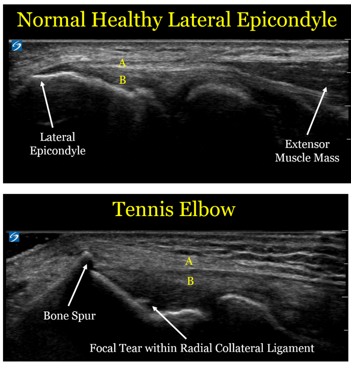
Here are comparison ultrasound images of a healthy lateral epicondyle, followed by the lateral epicondyle of a patient with tennis elbow. 


Let's look at the ultrasound images above, first focusing on the upper image of a healthy tendon.  One useful thing to see visually is that the muscle mass (the right arrow) looks distinctly different than the extensor tendon (designated by the yellow letter "A").  This is an important detail, since muscle has an excellent blood supply and heals well.  Tendon is made of a tight bundle of collagen with a relatively poor blood supply, and may struggle to heal.

In the second image, we can see the anatomic changes associated with tennis elbow:
1. The extensor tendon (A) is thickened and not as well defined as in the healthy elbow.  Of note, the patient with the healthy tendon is actually a much larger and more muscular person, so the thickened tendon is in fact a sign of an unhealthy tendon
2. The bone spur created by the unhealthy tendon.  We call this an enthesophyte, which is where hte tendon inserts onto the bone.  The reason these enthesophytes form is because bone is really a very dense putty, so the enthesophyte is caused by abnormal pulling forces by the unhealthy tendon
3. The part labeled "B" is the radial collateral ligament (RCL), which is underneath the extensor tendon.  In the case of the unhealthy patient, there is a tear in the RCL.  By definition, a sprain is a tear in a ligament.
4. Of note, there is no "inflammation", which is why the term -itis (e.g., tendonitis) is usually a misnomer.  This is an example of a chronic degradation of the extensor tendons and underlying radial collateral ligament, and the more proper term would be tendinosis or tendinopathy.


For a sake of comparison, the image below is an MRI


The level of detail is far less.  The resolution (which is defined as the ability to distinguish 2 points as distinct) is 5 times higher for the ultrasound.  In the ultrasound image, there is a clear distinction between the extensor tendons and the underlying radial collateral ligament, and we can see the individual fibers with great detail.  In the MRI, the white arrows show what is essentially a black line, with far less detail.  MRI does have some advantages in other contexts (e.g., it sees a wider field of view and can penetrate the bone, so it helps with bone contusions and certain types of joint injuries), but for imaging the lateral epicondyle, ultrasound is a better choice.


So what should we do with these patients?
- Historically, physicians have often performed landmark-based injections to the lateral epicondyle of cortisone.  However, moreso than for any other sports injury, repeated data show this is not a good idea.  While cortisone injections may help in the short term, they clearly make the injuries worse in the long term.
- Similarly, patients are often advised to use the non-steroidal anti-inflammatory drugs (NSAIDs) like ibuprofen or naproxen.  However, these are also bad choices, because while they can help with pain in the short term, they also prevent healthy collagen formation.
- The best initial treatment option is repeated icing.  Ice is a better anti-inflammatory than any medication, and also has other healing benefits.
- Physical therapy is also a mainstay of the best treatment for tennis elbow, where they can focus on exercise protocols designed to promote healing and fix damaging biomechanical patterns.
- For the subset of patients who do not improve with either ice or physical therapy, we often recommend use of proliferative therapy techniques including platelet-rich plasma injections or prolotherapy, which can help induce healthy healing.